Histology for Healthy Skin:: Layers, Functions, and Clinical Importance
Histology for Healthy Skin: A Complete Guide
Introduction
The skin is far more than just a surface that defines our appearance—it is the body’s largest organ and its first protective shield against the outside world. It guards us from environmental hazards, regulates temperature, allows us to sense the world, and even plays a role in immunity and vitamin production. To truly understand how skin maintains its beauty, strength, and resilience, we need to look beyond what the eye can see and explore it under the microscope.
This is where histology for healthy skin—the study of tissues—becomes essential. Histology for Healthy Skin helps scientists, dermatologists, and skincare experts uncover the microscopic architecture of the skin. Through this science, we can see how skin renews itself, why it ages, and how diseases alter its structure. In short, Histology for Healthy Skin provides the foundation for dermatology, cosmetic medicine, and skincare innovation.
The Vital Functions of Skin
The skin is not just a covering but a dynamic organ with life-sustaining roles. Its functions can be grouped as follows:
- Barrier Protection—Prevents harmful bacteria, viruses, toxins, and ultraviolet (UV) rays from penetrating the body while reducing water loss.
- Thermoregulation—Sweat glands release fluid to cool the body, while blood vessels widen or narrow to control heat flow.
- Sensation—Specialized nerve endings detect touch, pain, vibration, and temperature, allowing us to interact with the world safely.
- Vitamin D Synthesis—Sunlight triggers chemical reactions in the skin that produce vitamin D, essential for bone and immune health.
- Immune Defense—Skin cells such as Langerhans cells detect harmful microbes and activate immune responses.
Each of these functions is closely tied to the skin’s microscopic design, which histology allows us to study in detail.
Tissue Preparation in Skin Histology
Before skin can be observed under a microscope, it must undergo tissue preparation, a process that preserves and reveals microscopic details:
- Fixation—Chemicals like formalin preserve tissues, preventing decay.
- Embedding—The sample is encased in paraffin wax to stabilize it.
- Sectioning—Ultra-thin slices (3–10 micrometers) are cut using a microtome.
- Staining—Dyes such as hematoxylin and eosin (H&E) highlight cell nuclei, cytoplasm, and connective structures.
This preparation transforms invisible structures into clear, colorful patterns that pathologists and researchers can analyze.
Advanced Staining in Histology for Healthy Skin: Histochemistry and Cytochemistry
While standard staining shows basic tissue structures, advanced techniques in histology for healthy skin reveal important biochemical details:
- Histochemistry—Uses chemical reactions to highlight molecules like glycogen, lipids, and collagen.
- Cytochemistry—Focuses on cellular components, such as enzymes and DNA, offering insights into cell activity.
In skin studies, these advanced staining methods within histology for healthy skin help identify melanin distribution, collagen quality, and keratin production—all of which are vital in diagnosing pigmentation issues, connective tissue disorders, and aging changes.
Microscopy in Skin Histology
Light Microscopy
- The most common technique, often paired with H&E staining.
- Reveals the basic epidermis, dermis, and hypodermis layers, along with hair follicles and glands.
- Widely used in diagnosing common conditions like eczema, psoriasis, fungal infections, and dermatitis.
Electron Microscopy
- Offers ultra-high magnification and resolution.
- Displays cellular connections like desmosomes, keratin filaments, and basement membranes in remarkable detail.
- Especially valuable in studying autoimmune diseases (e.g., pemphigus vulgaris) and genetic skin disorders.
Pathophysiology of Skin Through Histology
Histology provides a microscopic lens into disease processes:
- Acne—Blockage of sebaceous glands and bacterial overgrowth.
- Psoriasis—Rapid epidermal turnover leading to thickened scales.
- Melanoma – Malignant transformation of melanocytes.
- Aging – Loss of collagen, thinning dermis, and reduced elasticity.
Each condition leaves a unique histological fingerprint, making histology critical for accurate diagnosis and treatment planning.
Clinical Significance of Skin Histology
Skin histology is not just academic—it has wide-reaching clinical applications:
- Biopsies—Identify cancers, infections, and chronic conditions.
- Cosmetic Dermatology – Guides treatments like microneedling, chemical peels, laser therapy, and dermal fillers.
- Pharmaceutical Testing—Ensures that skincare and medical products are safe and effective.
- Forensic Medicine – Analyzes wounds, burns, or environmental exposure to aid legal investigations.
Thus, histology bridges scientific knowledge with real-world medical and cosmetic practice.
Layers of the Skin
In Histology for Healthy Skin, the structure of the skin is divided into three primary layers, each with specialized roles:
Epidermis—The outermost protective barrier made of stratified squamous epithelium.
Dermis—The middle layer rich in collagen, blood vessels, and nerves; provides elasticity and nourishment.
Hypodermis (Subcutaneous Tissue)—The deeper layer of fat and connective tissue that cushions and insulates the body.
Understanding these layers through histology for healthy skin helps explain how the skin maintains its strength, flexibility, and protection.
The Epidermis
Although avascular (without blood supply), the epidermis thrives by receiving nutrients from the dermis. Its main cell types include
- Keratinocytes—Produce keratin, giving skin strength.
- Melanocytes—Produce melanin for UV protection.
- Langerhans Cells—Provide immune surveillance.
- Merkel Cells—A—Provide—Act as touch receptors.
Stratum Basale & Stratum Spinosum
- Stratum Basale—Contains—Act stem cells that continually divide, fueling skin regeneration.
- Stratum Spinosum—Strengthened—Contains desmosomes, which link cells and provide resilience.
Stratum Granulosum & Stratum Corneum
- Stratum Granulosum—Produces keratohyalin granules, precursors of keratin.
- Stratum Corneum—Outermost layer of flat, dead keratinized cells that form a waterproof shield.
Melanocytes and UV Protection
Melanocytes, located in the stratum basale, produce melanin, the pigment that absorbs UV rays and prevents DNA damage. Variations in melanin production explain differences in:
- Skin color
- Tanning ability
- Susceptibility to skin cancer
Variations in Skin Thickness
Histology shows that skin thickness varies depending on location:
- Thick Skin—Found on palms and soles, with a thick stratum corneum; lacks hair follicles and sebaceous glands.
- Thin Skin—Covers most of the body, contains hair follicles and sebaceous glands, and is more flexible.
Special Structures of the Skin
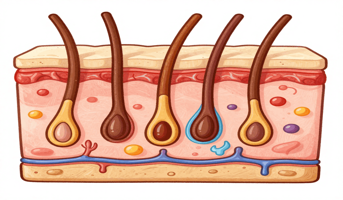
Hair
- Grows from follicles extending into the dermis.
- Composed of a bulb, shaft, and follicle wall.
- Provides protection, sensation, and aesthetic value.
Eccrine Sweat Glands
- Distributed across most of the body.
- Secrete watery sweat directly onto the skin surface.
- Essential for cooling and waste removal.
Apocrine Sweat Glands
- Found in armpits and groin.
- Release thicker secretions that produce odor when broken down by bacteria.
- Become active at puberty.
Sebaceous Glands
- Release sebum, a greasy material that lubricates hair and skin.
- Protects against dryness and bacterial invasion.
- Overactivity contributes to acne formation.
Sensory Structures in Skin
The skin is highly sensitive thanks to specialized receptors:
- Meissner’s Corpuscles – Detect light touch.
- Pacinian Corpuscles—Respond to deep pressure and vibration.
- Free Nerve Endings – Sense pain, heat, and cold.
These structures enable the skin to act as a sensory organ of communication with the environment.
Review Questions with Answers
Q1. What are the three main layers of skin and their functions?
- Epidermis – Outer barrier protecting against UV rays, pathogens, and water loss.
- Dermis—Provides elasticity, strength, and nourishment.
- Hypodermis – Cushions the body, stores fat, and insulates.
Q2. How do melanocytes protect skin from UV rays?
Melanocytes in the stratum basale produce melanin, which absorbs UV radiation, prevents DNA mutations, and lowers cancer risk.
Q3. Why are sebaceous glands important?
They secrete sebum, which hydrates skin, maintains softness, provides mild antibacterial protection, and waterproofs the skin.
Q4. Why is histology vital in diagnosing skin diseases?
Because microscopic examination reveals disease-specific changes, allowing accurate diagnosis of conditions like psoriasis, melanoma, eczema, and infections.
Q5. What is the difference between thick and thin skin?
- Thick skin (palms, soles) has a thicker stratum corneum, no sebaceous glands, and no hair.
- Thin skin (rest of the body) has hair follicles and sebaceous glands and is more flexible.
Conclusion
Histology for Healthy Skin offers a microscopic roadmap to understanding how the skin maintains its beauty, strength, and protection. From the epidermis to the dermis and hypodermis, each layer contributes to resilience, immunity, and sensory function. Structures like hair, glands, and nerve endings further enrich the skin’s role as both a shield and a communicator with the outside world.
By studying histology for healthy skin, scientists and dermatologists can diagnose diseases more accurately, develop innovative treatments, and guide people toward healthier skin.
Ultimately, the deeper we explore the skin’s microscopic architecture, the better we can preserve its vitality, radiance, and function throughout life.
Disclaimer:
This article’s product reviews, routines, and skincare advice are provided solely for informational purposes. Each person’s skin type—oily, dry, or sensitive—reacts differently to substances like vitamin C, retinol, and homemade masks. Before attempting any new product or solution, we highly advise conducting a patch test. Please see a licensed dermatologist if you have ongoing skin issues or before beginning clinical therapies. Individual outcomes might differ.


8 Comments